


- home
- >
- 過去のセミナーでの症例
- >
- 2011年度症例
Case Presentation at Endo-Skill Update 2011
ESU2011にてご提示した症例(※症例画像を許可なく複製、使用することを禁じます。)
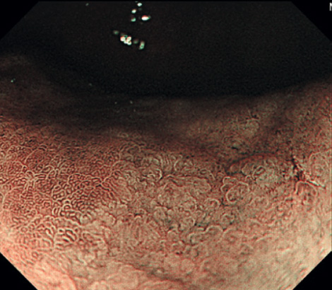
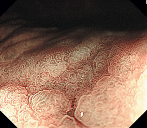
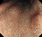
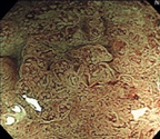
case1A gastric depressed lesion
A shallow depressed lesion was shown in the greater curvature of the mid-gastric body. The demarcation of this lesion was unclear in white light. NBI magnified endoscopy showed irregular surface and vascular pattern. The demarcation was diagnosed clearly by NBI magnified endoscopy. How do you diagnose this lesion?
No.1
No.2
No.3
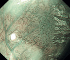
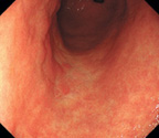
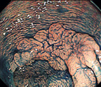
case2An esophageal depressed lesion
A reddish shallow depressed lesion was shown on the left wall of the middle esophagus. NBI endoscopy revealed a brownish area. NBI magnified endoscopy showed irregular micro vascular pattern. How do you diagnose this lesion?
No.1
No.2
No.3
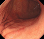
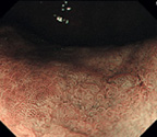
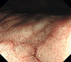
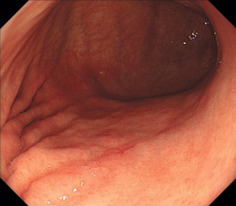
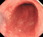
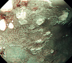
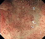
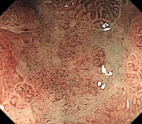
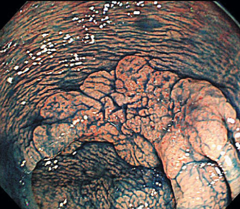
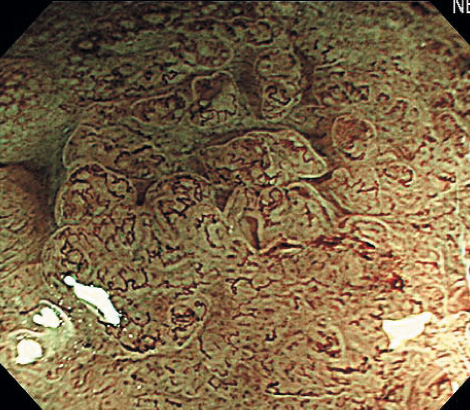
case3A gastric depressed lesion.
A large shallow depressed lesion was found in the antrum. The color of the lesion wasn`t homogeneous, and the margin of the lesion was indistinct. NBI magnified endoscopy revealed irregular villous or unclear surface pattern. And irregular vascular pattern was observed in the unclear area. The size of the lesion is 2cm or more. Therefore, diagnosis of the histology is important to decide the treatment strategy. How do you diagnose this lesion?
No.1
No.2
No.3

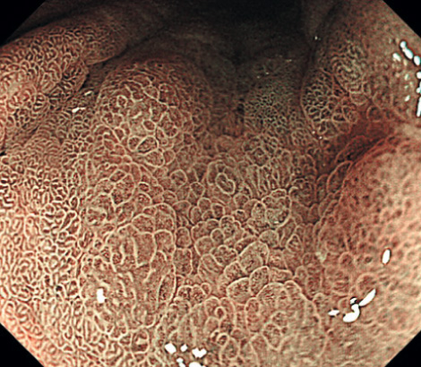
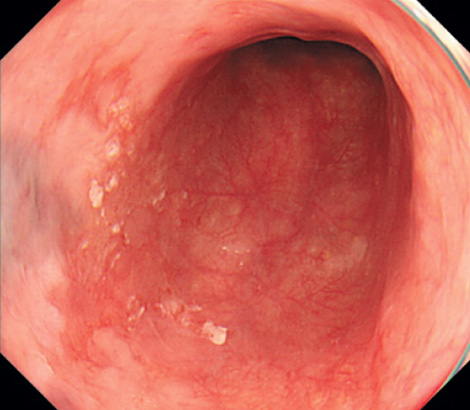
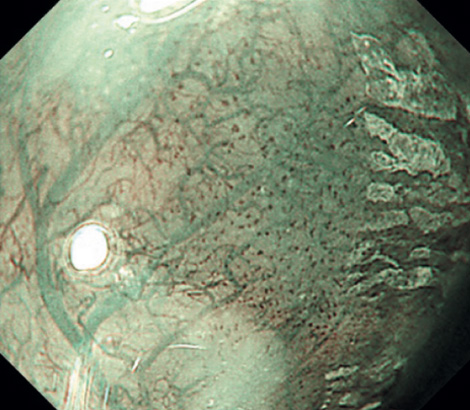
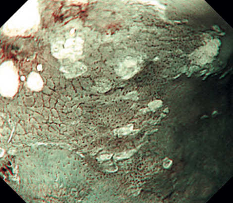
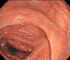
case4A colonic flat elevated lesion.
A flat elevated lesion existed in the transverse colon. There was a shallow depressed area with mucosal contraction in the center part of the lesion. NBI magnified endoscopy revealed irregular surface and vascular pattern. How do you diagnose this lesion? Do you treat this lesion by EMR, ESD or Colectomy?
No.1
No.2
No.3

ABOUT
ESUについて
本研究会(エンド スキル アップデート)では最先端の内視鏡診断に関するライブデモンストレーションを行い、最新の診断手技、そしてESDのコツとポイントを紹介します。
-
This year's seminar
ESU2016で
提示予定の症例 -
Endo-Skill Update
2016

-
Past Seminars
過去のセミナーでの症例 -

Endo-Skill Update
2015
-

Endo-Skill Update
2014 -

Endo-Skill Update
2013 -

Endo-Skill Update
2012 -

Endo-Skill Update
2011 -

Endo-Skill Update
2010 -

Endo-Skill Update
2009 -

Endo-Skill Update
2008